|
The spinal cord is a thin column of
nerve tissue that extends downward from the brain
through the vertebral column to the level of the second
lumbar vertebra. The spinal cord transmits pain signals
and other nerve impulses to and from the brain and
controls reflex actions. The spinal cord is
approximately 18 inches (46 cm) in length and up to 0.75
inches (1.9 cm) in diameter at its widest point.
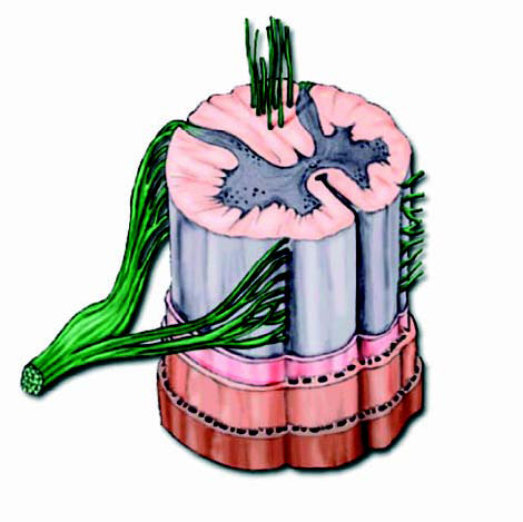
The spinal cord has three main components:
● Spinal nerves
● Nerve tissue
● Meninges |
|
|
Spinal
nerves
The spinal nerves originate in the spinal cord and carry
impulses to muscles and other structures, and return
impulses received from sensory organs. There are 31
pairs of spinal nerves that provide two-way transmission
of nerve impulses between the spinal cord and the arms,
legs, neck, and trunk.
Spinal nerves contain both sensory (incoming) and motor
(outgoing) nerve fibers. The sensory nerve fibers enter
the spinal cord from the dorsal (back)
area. The motor nerve fibers exit through the anterior
(ventral) area.
Each spinal nerve pair emerges from the spinal cord from
two short branches, or roots, which lie within the
vertebral column:
● Dorsal root
● Ventral root |
|
|
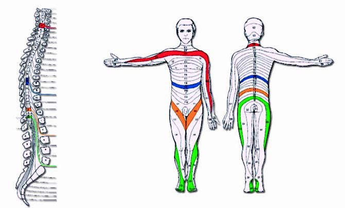
Dermatomes
A dermatome is an area of the body supplied by a
particular section of spinal nerves connected to a
specific vertebral body. In the torso, the dermatomes
form consecutive bands. A map of these bands, called a
dermatome map, is an important diagnostic tool for pain
syndromes. |
|
|
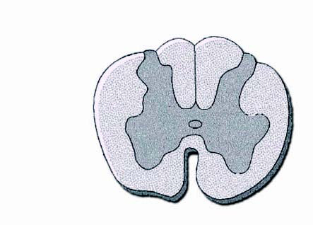
Nerve tissue
The spinal cord consists of two types of nerve tissue:
● Grey matter
● White matter |
|
|
Grey matter
Grey matter makes up the centre of the spinal cord. Grey
matter consists primarily of dendrites, which are
thread-like extensions of a neuron, and cell bodies of
neurons. Both of these neuron components receive nerve
impulses from other neurons. |
|
|
White matter
White matter surrounds the grey matter. White matter
consists of bundles of nerve fibers that form conduction
paths called spinal tracts. The nerve fibers are
surrounded by myelin. Nerves that are myelinated
transmit information to and from the brain faster than
nerves that are not myelinated. |
|
|
Meninges
Meninges are membranous tissue coverings that surround
and protect the spinal cord from injury. There are three
types of meninges that surround the
spinal cord:
● Dura mater
● Arachnoid membrane
● Pia mater
Dura matter is the tough, protective, outer covering
that is made up of collagenous (protein) tissue. The
arachnoid membrane is a delicate membrane that covers
the subarachnoid, or intrathecal space. The pia matter
is a vascular membrane that closely attaches to the
spinal cord and surface of the brain. The pia matter is
the innermost layer of the meninges. |
|
|
Intraspinal
space
The layering of the dura matter, arachnoid membrane and
pia matter creates ‘spaces’ in the spinal cord, known as
intraspinal spaces. There are two types of intraspinal
spaces:
● Epidural space
● Intrathecal or subdural space |
|
|
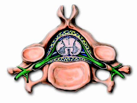
Epidural
space
The epidural space lies between the vertebrae and the
dura matter. The epidural space is filled with fatty
tissue and consists of a network of large, thin-walled
veins and spinal nerve extensions. This is shown in
Figure 3.Neurostimulation leads are placed in the
epidural space. |
|
|
Intrathecal
or subdural space
The intrathecal or subdural space is a potential space
that is located between the dura mater and the arachnoid
membrane. This small-volume space is filled with
cerebral spinal fluid (CSF). CSF completely bathes the
brain and spinal cord, which provides a fluid cushion
against shocks. This is shown in Figure 6. The drug
delivery catheter is usually placed in the intrathecal
space to provide site-specific delivery of a
pain-relieving drug such as morphine to opioid receptors
in the spinal cord. However the screening test (which
tests a patients response to intraspinal morphine) can
often be conducted by delivering the drug to the
epidural space).
|
 What’s Up